Anatomy of a Medical Term:
•Prefix: word part found at
the beginning of a word
Prefixes usually, but not
always, indicate location, time, number, or status.
Prefixes are presented
with a hyphen following them: prefix-
•Pre- means before:
–Preoperative means before an
operation.
•Peri- means around:
–Perioperative means pertaining to
the time around an operation.
•Post- means after:
–Postoperative means after an
operation.
•Root: word part that gives
the essential meaning of the word
Roots are the foundation
of medical terms.
Roots usually, but not
always, describe the part of the body that is involved.
Roots cannot stand alone:
–A suffix must be added to complete the term.
–A prefix may be added to the term.
•Suffix: word part found at
the end of a word
•Suffixes usually, but not
always, indicate the procedure, condition, disorder, or disease.
•There are many related
groups of suffixes:
–“pertaining to” suffixes
–surgical suffixes
–procedural suffixes
–the double r
suffixes
–conditional and structural suffixes
•Suffixes
may change the part of speech of a word.
•Different
suffixes may change the word from a noun to an adjective.
•Combining vowel: a single
vowel, usually an o, that is added to the end of a root to make
the word easier to pronounce
•A combining vowel may be
used to make the medical term easier to pronounce.
•A combining vowel is not
used when the suffix begins with a vowel.
•A combining vowel is used
when the suffix begins with a consonant.
•A combining vowel is used
when two or more word roots are joined.
•A prefix does not need a
combining vowel.
•Combining form: the
combination of the root and the combining vowel
•Combining forms = root
plus a vowel
•There may be more than one
combining form for a body part.
•Combining forms will be
presented with a backslash: hepat/o.
•Examples of combining
forms:
–Nas/o means nose.
–Rhin/o means nose.
–Ren/o means kidney.
–Nephr/o means kidney.
Public Health and Ethics
—Zoonosis:
—zoonoses;
disease
of animals transmissible to humans. Saunders
Comprehensive Veterinary Dictionary
What kind of
diseases?
Rabies,
brucellosis, tuberculosis, anthrax, E. coli, Salmonella
Sanitation: —The establishment of
conditions favorable to health, especially with respect to infectious diseases.
- Antiseptics- generally used on living tissue
- Disinfectants- generally used on objects
- These are concentration dependent. Some things full concentration are good for a disinfectant but have to be diluted to be used as a antiseptic. EX- iodine on a calf's navel at full strength can damage the living tissue but diluted it is good.
- Disinfection- is the destruction of the vegetative forms of bacteria
- vegetative- living and growing bacteria
- It is important to note that when they perform a surgical scrub on the patients skin they are not sterilizing it. They are only disinfecting it/ lowering the bacterial numbers
- Sterilization- is the destruction of ALL organisms and spores on an object to stop disease (bacteria, virus, fungus, spores)
Physical Sterilization:
- Filtration- commonly used for liquids and gases- Pharmaceuticals are commonly sterilized by filtration
- Radiation- why radiation over heat? because things like surgical gloves will melt in heat- heat sensitive
- Heat- an object is not sterilized until the steam has permeated the whole pack (autoclave) at the proper time, temperature, and pressure.
Chemical Sterilization:
- Is used on heat sensitive things like plastic--most common is ethylene oxide
What is the Gold Standard? Put a vile of bacteria in the pack going into the autoclave. Run as directed. Remove and collect a swab and then give it time and see if it grows. This method is not very practical or do able.
What is cold sterile? Not technically sterile but it is when the tools/instruments are submerged in a disinfectant. Used for non-sterile procedures.
What is aseptic technique? A way of doing things without microorganisms. Gown up/glove up.
a- without or not
sepsis- presence in the blood or other tissues of pathogenic microorganisms or other toxins
What is a pathogen? It is bacteria, virus, fungus
Practice Act—
—Limitations
on Supervising Veterinarians. Unless otherwise provided by law or rule, a
supervising veterinarian shall not authorize a certified veterinary technician,
a veterinary technician working under a temporary certification, an assistant
or anyone else, other than a licensed veterinarian or a veterinarian holding a
valid temporary permit to perform the following functions:
—a. Surgery;
—b. Diagnosis and
prognosis of animal disease;
—c. Prescribing drugs,
medicines and appliances; or
—d. Diagnosis and
performance of procedures that constitute operative dentistry/oral surgery as
defined by Section 54-2103(13)(b), Idaho Code.
"Direct
supervision"
—means the supervising
veterinarian is on the premises where the animal is being treated, is quickly
and easily available and the animal has been examined by the supervising
veterinarian as acceptable veterinary medical practice requires.
—"Immediate
supervision"
—means the supervising
veterinarian is in the immediate area, in audible and visual range of the
animal patient and the person treating the patient and the animal has been
examined by the supervising veterinarian as acceptable veterinary medical
practice requires.
—"Indirect
supervision"
—means the supervising
veterinarian is not on the premises but is available for immediate contact by
telephone, radio or other means, has given either written or oral instructions
for treatment of the animal patient, the animal has been examined by the
supervising veterinarian as acceptable veterinary medical practice requires,
and the animal, if previously anesthetized, has recovered to the point of being
conscious and sternal.
VCPR:
—Veterinary-Client-Patient
Relationship
—When does this exist?
1.The
veterinarian has assumed responsibility for making clinical judgments regarding
the health of the animal(s) and the need for medical treatment, and the client
has agreed to follow the veterinarians instructions.
2.The
veterinarian has sufficient knowledge of the animal(s) to initiate at least a
general or preliminary diagnosis of the medical condition of the animal(s).
This means that the veterinarian has recently seen and is personally acquainted
with the keeping and care of the animal(s) by virtue of an examination of the
animal(s), or by medically appropriate and timely visits to the premises where
the animal(s) are kept.
3.The
veterinarian is readily available, or has arranged for emergency coverage, for
follow-up evaluation in the event of adverse reactions or the failure of the
treatment regimen.
Anatomy- the study of body structure
Physiology- the study of body function
Pathology- the study of disease
Etiology- the study of the cause of disease
Cytology- the study of cells
Histology- the study of tissue
Directional Terms:
Medial- towards midline
Lateral- away from midline
Cranial- towards the head
Caudal- towards the tail
Proximal- closer to the body, nearest to the beginning of a structure
Distal- farther from the body, farthest from the beginning of the structure
Anterior- towards the head
Posterior- towards the tail
Superior- above, on top
Inferior- below, lower
Superficial- on top, outside, near the surface
Deep- away from the surface, internal
Adduction- movement towards the midline (think of adding something)
Abduction- movement away from the midline (think of child abduction)
Flexion- angle gets smaller, decreases
Extension- angle gets bigger, increases
Rostral- towards the nose end of the head
Cephalic- pertaining to the head
Body Planes:
Median/midsagittal- divides the body into equal left and right halves
Sigittal- unequal left and right parts
Dorsal- back plane
Ventral- belly plane
Transverse- perpendicular to the long axis
Body Cavities:
Cranial
Spinal
Thoracic
Abdominal
Pelvic
Recumbency:
Whatever aspect is contacting the ground
Dorsal- on the back
Ventral- on the belly
Lateral- on the side (left/right)


Common Anatomical Terms:


Musculoskeletal System:
The musculoskeletal system
consists of two systems that work together to support the body and allow for movement of the animal:
–the skeletal system = bones, joints, tendons, cartilage, ligaments, and various
connective tissues
–the muscular system = muscles and various connective tissues
Bones- oste/o, oss/e, oss/i:
1. form the framework that supports and protects the body
2. aid in body movement
3. function to form blood cells
4. store and release minerals such as calcium
Joints- arthr/o:
Connections between bones
Aid in movement of the body
Types of Joints:
Synarthroses- immovable- skull sutures
Amphiarthroses- slightly movable- pelvic symphysis joint
Diarthroses- freely movable- shoulder, hip, knee joint
Cartilage- chondr/o:
Cartilage is a form of connective tissue that is more elastic than bone
Protects the ends of the bones where they contact each other
Ligaments- ligament/o:
Bands of fibrous connective tissue that connect one bone to another
Tendons- ten/o, tend/o, tendin/o:
Bands of fibrous connective tissue that connect muscle to bone
Skeleton:
The skeleton is divided into two parts:
- Axial Skeleton- protects the major organs of the nervous, respiratory, and circulatory systems
- It consists of the skull, hyoid bones, vertebral column, ribs, and sternum
- Appendicular Skeleton- the framework that consists of the extremities, shoulder, and pelvic girdle
- Appen means to add or hang
- It includes the bones of the front and hind limbs
Types of Bones:
- Long
- Femur
- Humerus
- Radius
- Ulna
- Short
- Carpal/tarsal bones
- Sesamoid= small bone embedded in a tendon or ligament
- horse sesamoid bones
- Flat bones
- bones of the skull
- Irregular bones
- Vertebrae
Structures of the Skeletal System
Bone Anatomy Terms:

- Diaphysis- the long shaft/body of the bone
- Epiphysis- the enlarged ends of the bone
- Metaphysis- joining portion of the diaphysis and epiphysis- the physis (growth plate) is in there
- Periosteum- lining around the bone- important for bone growth, repair, nutrition, attachment of tendons and ligaments
- Articular Surface- where one bone comes in contact with or articulates with another. This is usually on the epiphysis and covered with hyaline cartilage (articular cartilage)
- Medullary Cavity- space in diaphysis where bone marrow is
- Endosteum- layer of tissue lining the medullary cavity
- Apophysis- any outgrowth of a bone or process
- Cortex or Cortical or Compact- hard, dense, strong bone-compact bone surrounding the medullary cavity
- Growth Plate- found in the metaphysis
- Compact Bone- refer to 9
- Cancellous Bone- lighter, less strong bone- lattice like structure
Thoracic Limb (lateral view) of a Dog:

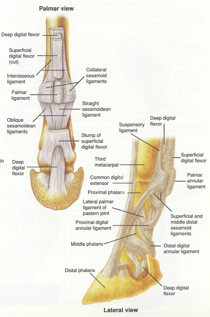
Thoracic Limb (lateral view) of a Horse:


Dog Digits:

Ox digits:

Horse digits:

Pelvic limb (lateral view) of a Dog:

Pelvic limb (lateral view) of a Horse:

The Muscular System:
Muscles are organs that contract to produce movement.
Muscles are responsible for the following:
- ambulation
- control of organs and tissues
- pumping of blood
- generation of heat
Muscles are made up of long, slender cells called muscle fibers.
Each muscle consists of a group of muscle fibers in a fibrous sheath.
- My/o- is the combining form for muscle
- Fibr/o and fibros/o- are combining forms for fibrous tissue
Types of Muscle Tissue:
- Skeletal Muscle
- voluntary= control
- striated
- Cardiac Muscle
- involuntary= no control
- heart muscle
- striated
- Smooth Muscle
- involuntary= no control
- arteries, intestines, stomach, uterus
- non-striated
Anal Sphincter= the smartest muscle in the body because it can determine the difference between gas, liquids, and solids.
The Digestive System:
Also known as the alimentary system, gastrointestinal system, and the GI system
The digestive system is basically a long, muscular tube that begins at the mouth and ends at the anus
Functions of the digestive system:
- intake and digestion of food and water
- absorption of nutrients
- elimination of solid wastes
Structures of the Digestive System:
- mouth or oral cavity- or/o and stomat/o
- lips- labi/o and cheil/o
- cheeks- bucc/o
- palate- palat/o
- tongue- gloss/o and lingu/o
- jaw- gnath/o
- prognathia means having an elongated mandible (overshot)
- brachygnathia means having a shortened mandible (undershot)
- teeth- dent/o, dent/i, and odont/o
- the primary dentition is temporary and known as the deciduous dentition
- decidu/o= shedding
- Types of teeth
- incisor- front cutting tooth
- canine- long, pointed bonelike tooth for grasping and tearing
- premolar- cheek tooth that grinds food
- molar- caudal cheek tooth that grinds food
- gingiva- gingiv/o
- salivary glands- sial/o and sialaden/o
- pharynx- pharyng/o
- joins the respiratory and gastrointestinal systems. AKA throat
- esophagus- esophag/o
- collapsible, muscular tube that leads from the oral cavity to the stomach. AKA gullet
- enters the stomach through an opening that is surrounded by a sphincter
- stomach- gastr/o
- saclike organ that aides in digestion of food
- animals can be classified as monogastric or ruminant
- monogastric animals have one true glandular stomach (one that produces secretions)
- ruminants have one true glandular stomach and three forestomachs
- ruminants regurgitate and remasticate their food
- rumen, reticulum, omasum, abdomasum
- cows, sheep, goats, deer have 4 and alpaca, llamas, camels only have 3
- an adult cows rumen holds about 50 gallons and the contractions stir the food counter clockwise if you are on the right side
- small intestines- enter/o
- extends from the pylorus to the large intestine. Held in place by the mesentery. Absorption occurs here. Villi are tiny hairlike projections that increase the surface area of the small intestine
- It has three segments
- duodenum- proximal part (duoden/i or duoden/o)
- jejumum- middle part (jenun/o)
- ileum- distal part (ile/o)
- large intestines
- extends from the ileum to the anus
- It has four segments
- cecum- cec/o
- colon- col/o
- rectum- rect/o
- anus- an/o or proct/o= anus and rectum together
- accessory organs of digestion
- Liver- hepat/o
- Gallbladder-
- chol/e= bile
- cyst/o= sac
- doch/o= receptacle
- Pancreas- pancreat/o
- Salivary glands
Peristalsis- series of wavelike contractions of smooth muscle that moves the ingesta through the tract
Segmentation- involves the side-to-side mixing of ingesta
Functions of the Urinary System:
- The urinary system removes waste from the body by constantly filtering the blood. In addition to filtering, the the urinary system also maintains proper balance of water, electrolytes, and acids in body fluids and removes excess fluids from the body. Maintaining a proper balance of water, electrolytes, and acids allows the body to have a stable internal environment. This stable internal environment is called homeostasis.
- The structures of the normal urinary system include a pair of kidneys, a pair of ureters, a single urinary bladder, and a single urethra.
- Urine is formed in the kidneys, flows through the ureters to the urinary bladder, is stored in the urinary bladder, and flows through the urethra and outside the body.
- Cortex and Medulla= outer and inner
- Hilus= nerves, blood supply, ureters
- Nephron= functional unit of the kidney
- Renal Pelvis= area where the urine collects before entering ureters (only present in some species)
- Glomerulus= cluster of capillaries
- Bowman's capsule= cup shaped structure that contains the glomerulus
- Cystocentesis- puncture the bladder and and draw out urine
Cardiovascular System:
Delivers oxygen, nutrients, and hormones to various tissues of the body.
Transports waste products to the appropriate waste removal system.
Is also referred to as the circulatory system.
Cardiovascular means pertaining to the heart and blood vessels.
The heart is a hollow muscular organ that provides the power to move blood through the body.
The heart is located in the mediastinum, a space in the thoracic cavity between the lungs.
The Structures Surrounding the Heart:
- The pericardium is a double-walled membrane that surrounds the heart
- Peri- means around
- The pericardium has two layers:
- fibrous layer
- serous layer
- parietal layer
- visceral layer
- The pericardial space is the space between the two serous layers of the pericardium.
- This space contains pericardial fluid
- Pericardial fluid prevents friction between the heart and the pericardium when the heart beats
Blood Supply to the Heart:
The blood vessels that deliver blood to and take blood away from the heart are known as coronary vessels.
-Coronary occlusion means blockage of coronary vessels.
-Coronary occlusion may lead to ischemia. Ischemia is a deficiency in blood supply to an area.
-Ischemia may lead to necrosis. An area of necrosis caused by an interrupted blood supply is an infarct. A myocardial infarct is a heart attack.
The Heart Chambers:
- The superior chambers of the heart are known as atria (singular is atrium) -atri/o
- The inferior chambers of the heart are known as ventricles- ventricul/o
- Septum- is a separating
- Apex- is the tip of the heart
The Heart Valves:
- a valve is a membranous fold
- heart valves control the flow of blood through the heart- valv/o and valvul/o
- Right Atrioventricular valve is also known as the tricuspid valve
- Pulmonary semilunar valve
- Left Atroventricular valve also known as the mitral valve and bicuspid valve
- Aortic semilunar valve
Heart Rate:
- Heartbeat= the rate and regularity of the heart rhythm
- Heartbeat is influenced by the electrical impulses from nerves that stimulate the myocardium
- Sinoatrial node (SA node) is located in the right atrial wall and initiates the heart rhythm
- it is called the pacemaker of the heart
- Atroventricular node (AV node) is located in the septum and receives impulses from the SA node
The Heart Walls:
- Epicardium- outside
- Myocardium- middle/muscle
- Endocardium- inside
Heart Rate Terms:
- Systole: contraction
- Asystole: without contraction
- Diastole: relaxation
- Arrhythmia: abnormal heart rhythm
Heart Rate Terms:
- Bradycardia: abnormally slow heartbeat
- Tachycardia: abnormally fast heartbeat
Electrocardiography- (ECG or EKG) is the record of electrical activity of the myocardium. This is the process of recording electrical activity of the heart.
Auscultation is listening to body sounds with a stethoscope.
When the heart is auscultated, a lubb/dubb sound is heard
-lubb= closing of the AV valve
-dubb= closing of the semilunar valve
-murmur= abnormal sound associated with turbulent blood flow
Blood Vessels- vas/o and angi/o:
- 3 Major types of blood vessels:
- arteries- arteri/o (high pressure)
- vessels that carry oxygenated blood away from the heart
- smaller arteries are arterioles
- arteries have no valves
- capillaries
- single-cell-thick vessels that connect the arterial and venous systems
- veins- ven/o and phleb/o (low pressure)
- vessels that carry deoxygenated blood toward the heart
- valves and muscles are what get the low pressured blood back to the heart
Blood pressure is the tension exerted by blood on the arterial walls
The two exceptions:
Pulmonary artery is not oxygenated- it is going to the lungs
Pulmonary vein is an oxygenated vein that is coming to the heart from the lungs
Red= oxygenated blood
Blue= deoxygenated blood
Vena Cava dumps into the---right atrium---right atrio ventricular valve---right ventricle---pulmonic valve-pulmonary artery---arterioles---capillaries (becomes oxygenated)---venules---pulmonary veins---left atrium---left AV valve---left ventricle---aortic valve---aorta---systemic arteries---arterioles---capillaries (goes from oxygenated to deoxygenated)---venules---vena cava
The Respiratory System:
The respiratory system brings oxygen from the air into the body for delivery via blood to the cells.
Respiration is the exchange of gases (oxygen and carbon dioxide) between the atmosphere and the body cells.
Ventilation means the bringing in of fresh air/ AKA breathing
The respiratory system is divided into upper and lower tracts
Upper Respiratory Tract:
- nose- air enters and exits the body through the nose
- mouth
- pharynx
- epiglottis
- larynx
Lower Respiratory Tract
- trachea
- bronchial tree
- bronchioles- no cartilage rings
- alveoli
- lungs- main organ of respiration- pneum/o, pneumon/o, pneu= means lungs or air and pulm/o and plumon/o mean lung
- the lung is encased in a membranous sac called the pleura
- the pleura has two layers; the parietal pleura and the visceral pleura
- between these layers is the pleural space
-pnea= means breathing
ox/i, ox/o, and ox/y= oxygen
capn/o= carbon dioxide
No comments:
New comments are not allowed.